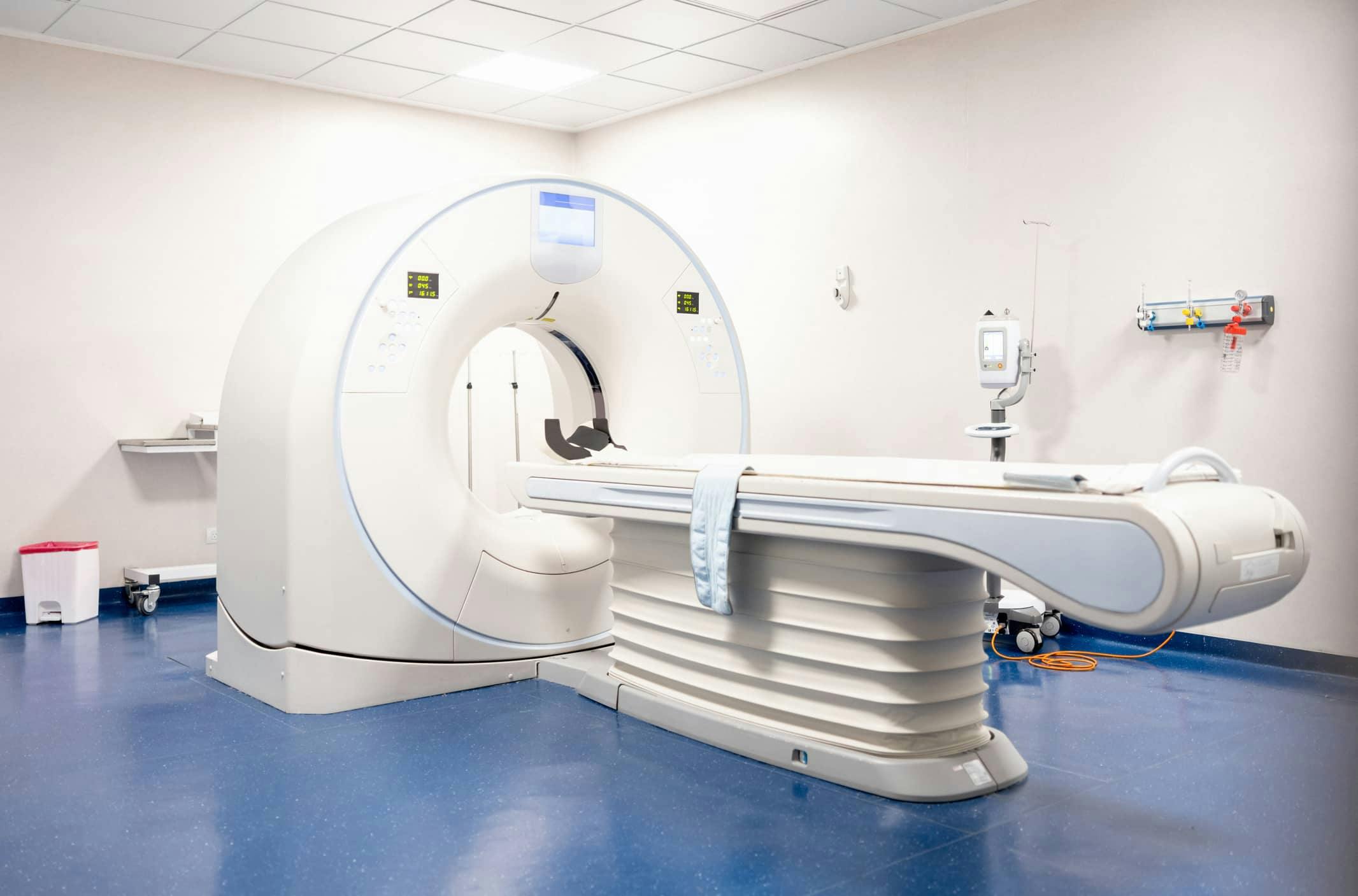
What is an MRI?
One of the most advanced diagnostic imaging methods available, the MRI provides detailed information about the body and is often used to evaluate the spine. Unlike plain X-rays, this non-invasive test not only shows the physician the bones, but also provides excellent details of the soft tissues. The MRI is helpful in diagnosing any condition in which the anatomy of the spine and its soft tissues need to be seen clearly, including degenerative disc disease, herniated disc, spinal stenosis, kyphosis, sciatica, scoliosis and tumors and infections of the vertebrae. However, since the MRI is more expensive and generally takes longer than the X-rays or CT scans, it is usually not the first test given.
Instead of X-rays, a MRI uses magnetic waves to take pictures of the spine. A magnet excites the hydrogen atoms in the body, which give off electromagnetic waves that are recorded by a computer. The computer then analyzes the results to reconstruct an image of the spine.
The MRI scanner is a large tube with a table passing through it. Some people worry about feeling closed in; however, the tube always remains open on both ends. You won’t feel any pain, but you will hear some loud noises during the exam. Depending on how many pictures are needed, the exam can take anywhere from 20 minutes to 90 minutes.
It is very important that you notify your physician if you have any metal in your body which cannot be removed, such as a pacemaker, aneurysm clips or prosthesis. You may not be able to have an MRI if you have such metal in your body. Leave your jewelry at home, and remove all metallic objects, such as hearing aids and dentures, before entering the scanning room. There are “open” and “closed” MRIs; unless you are claustrophobic, a closed MRI is always performed.


