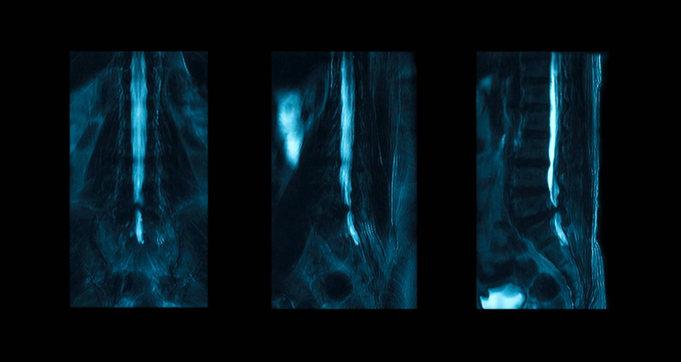
What is a Myelogram?
A myelogram is more invasive than a MRI, but is sometimes necessary to more accurately diagnose the patient’s condition. A CT scan is often performed at the same time as the myelogram in order to gather even more information. During a myelogram, special dye that shows up on X-rays is injected into the spinal column. The X-rays are then compiled into an image of the flow of fluid around your spinal column. Any decrease or blockage in the flow may indicate pressure on the nerves of the spine, possibly from a herniated disc or bony spur, or, less often, a tumor.
Administering the test involves lying on a X-ray table, where you are given a local anesthetic to numb the area. Then, a needle is placed into the area near your spine, dye is injected and finally the X-rays are taken. The test will take about an hour to complete. Afterwards, you may be taken for a CT scan, which can provide additional information that the myelogram may not find. For example, if a piece of disc has broken off and is pressing on a nerve somewhere other than its root, a CT-scan may detect it.
You won’t be able to eat solid foods after midnight the night before your test, but please drink plenty of fluids such as water or juice in order to be well hydrated. However, you will need to stop drinking fluids three hours beforehand. Ask your physician about any medications you may be taking. After your myelogram, you will need plenty of rest, and you cannot drive yourself home.
The doctor will discuss the risks of the myelogram, which are small but include itching around the puncture site, infection and allergic reactions. You may develop a headache either several hours or several days after the test. This is usually caused by a change in the pressure of the cerebral spinal fluid and should be cured with rest and plenty of fluids.


